Mesothelioma Diagnosis

Diagnosing mesothelioma is extremely difficult.
Because of this, many patients often endure months of pain and shortness of breath before they are properly diagnosed. According to an article published in Orphanet Journal of Rare Diseases, some patients are not diagnosed until they exhibit life-threatening complications, leaving them with limited treatment options.
The symptoms of mesothelioma are often mistaken for the flu, pneumonia or other respiratory illnesses. Unless the doctor is aware the patient had been previously exposed to asbestos, he or she may not consider malignant mesothelioma as a possible cause.
One of the reasons this disease is difficult to treat is that it is usually diagnosed in the late stages.
Early detection offers physicians a wider range of treatment options, including surgery, which are often not available options once the cancer has metastasized.
How can Mesothelioma be Diagnosed Early?
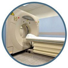
Mesothelioma Diagnostic Imaging Technologies
If your symptoms suggest mesothelioma your doctor will want to follow up with testing, beginning with imaging tests that allow your body to be viewed from the inside and checked for abnormalities such as the presence of fluid or tumors.
In addition to helping your doctor make an initial mesothelioma diagnosis, imaging technologies may be used for cancer staging, determining a treatment plan, and tracking response to treatments.

X-Ray Imaging: An abdominal or chest X-ray is typically the initial screening test for mesothelioma diagnosis. While they may show fluid collecting around the lung, abdomen or heart, thickening of the pleura, or hint at the existence of a tumor, X-rays don’t have enough resolution to identify the cancer and thus have limited diagnostic usefulness.

Computed Tomography (CT): A CT scan uses a series of X-rays to create three-dimensional computer images of the inside of the body that are better and more detailed than those created by traditional X-rays. CT scans are useful for gauging the location, characteristic and extent of the cancer. They may also be used to determine the viability of surgical treatment and the effects of treatment such as chemotherapy on the cancer. A CT scan by itself, however, cannot provide unequivocal mesothelioma diagnosis, although a CT guided biopsy—which uses a CT scan to guide the biopsy needle into the tumor—can.

Magnetic Resonance Imaging (MRI): An MRI scan creates detailed images of the body’s soft tissues using radio waves and magnets. Similar to a CT scan, MRI cannot unequivocally diagnose mesothelioma, but it is superior to a CT scan in terms of determining whether the cancer has metastasized. MRI is also useful for determining whether the tumor can be removed by surgery. Not all doctors use MRI for mesothelioma diagnosis. Those who do may use it in combination with CT scans for a more complete view of possible cancer sites.

Positron Emission Tomography (PET): Positron Emission Tomography scans measure functional and metabolic activity in the body through the use of radioactive glucose that’s injected into the bloodstream. The PET scan measures radioactivity in the body at certain “hot spots” that include the brain, heart and, in cancer patients, tumors. When a patient has a mesothelioma diagnosis, hot spots can indicate whether the cancer has spread to other areas.

PET-CT (Integrated PET – Computed Tomography): The PET-CT fusion scan combines the more-detailed CT scan with the biological activity-sensing PET scan into a single, more accurate image of what’s happening inside of the body. For example, the CT portion of the image may indicate an abnormality while the PET portion can show whether the abnormality is a metabolic “hot spot” indicative of malignancy, or rather an area of normal activity that suggests benignity.
Mesothelioma Diagnosis Process
Biopsy Procedures
Biopsy procedures remove tissue or cell samples for laboratory analysis. If you’re experiencing signs and symptoms of mesothelioma, your doctor may order a biopsy to determine whether you in fact have a mesothelioma diagnosis.
Biopsy procedures include:
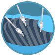
Thoracoscopy: During a thoracoscopy (a pleural biopsy), you’ll be put under general anesthesia and several small incisions will be made in your side. Through one of the holes will be inserted a thoracoscope (a thin, flexible tube like instrument with a light and camera attached) that allows your lungs to be seen on a video monitor. Through the other incisions, surgical tools are inserted and used to collect tissue samples that are then looked at under a microscope.

Laparoscopy: A laparoscopic biopsy is a way to collect a tissue sample when it’s suspected that a patient has peritoneal mesothelioma diagnosis. After the patient is put under general anesthesia, the surgeon makes small incisions in the abdominal wall. A laparoscope (a long, thin tube with a light and camera at the end) that allows the physician to see the peritoneal area on a monitor is inserted through one of the incisions. With a clear view of the region, biopsy samples are obtained and sent for laboratory analysis.

Bronchoscopy: A bronchoscopy does not involve making any incisions in the patient’s body. Rather, a flexible lighted tube with a camera (bronchoscope) is inserted down into the lung region through the mouth and used to look for abnormal areas in the airways. The bronchoscope may also have a tool to remove tissue samples for testing. Because this type of cancer doesn’t develop in the airways, a bronchoscope with an ultrasound device is used to find out if the cancer has spread into the lymph nodes in a procedure known as endobronchial ultrasound needle biopsy.
Mediastinoscopy: A mediastinoscopy is a procedure to check for cancer that’s spread to lymph nodes in the mediastinum, an area in the center of your chest. The procedure involves making a small incision at the base of the neck and examining the mediastinum with a special type of endoscope. Special instruments are then used to collect tissue samples for testing.
Diagnosis Statistics
Improvements in current treatments have provided patients with a more favorable life expectancy for people now being diagnosed with mesothelioma. Diagnosing the type and the stage of a person’s cancer is in, is very important to determine the patients life expectancy.
Many factors can affect survival, such as a person’s age and overall health, the treatment received, and how well the cancer responds to treatment.
The American Cancer Society provides some statistics about survival rates on patients diagnosed with mesothelioma.
Life Expectancy

Treatment after Diagnosis
After mesothelioma diagnosis has been confirmed, the doctor should explain the treatment options available to the patient.
There is no standard treatment, the doctor should plan a specific treatment each patient.
The most common treatment options involve:
- Surgery
- Chemotherapy
- Radiation
- Combination of these treatments called multimodal treatment plan.
Frequently Asked Questions
Can mesothelioma be misdiagnosed?
Because mesothelioma is a rare cancer, doctors may not immediately recognize symptoms. Patients may be told they have pneumonia or other lung conditions. It is important to discuss your history of asbestos exposure with your doctor. And do not hesitate to seek a second opinion from a mesothelioma specialist if you are experiencing symptoms.
How is mesothelioma diagnosed?
Malignant Mesothelioma is not easily diagnosed, further complicating treatment of a disease that is fast-spreading and resilient.
One of the first things patients experience is a dry cough and/or shortness of breath caused by a pleural effusion. Shortness of breath is experienced because the effusion (water around the lung) is pushing on the lung, making it difficult to breathe. Most patients presented with these symptoms receive initial treatment but the symptoms tend not to go away, or they may go away but only for a short amount of time.
When symptoms reappear after a few months, the cancer may already be advanced stage. Certain factors—such as a young patient, the absence of lung masses, or lack of a clear reason why the patient has a pleural effusion—are highly suspicious and warrant a more aggressive diagnosis. The earlier mesothelioma is diagnosed, the more varied the treatment options and the better the patient prognosis in most cases.
Most hospitals perform a thoracentesis (drainage of the lung with a needle), but 50 percent of these tests return a negative result. A more aggressive diagnostic method approach is therefore recommended, such as a biopsy or a pleuroscopy (camera-assisted biopsy). Diagnostic imaging techniques (i.e. X-rays, CT scans, and MRI) may assist with diagnosis but only a tissue sample can unequivocally diagnose mesothelioma.
Malignant Mesothelioma is not easily diagnosed, further complicating treatment of a disease that is fast-spreading and resilient. One of the first things patients experience is a dry cough and/or shortness of breath caused by a pleural effusion. Shortness of breath is experienced because the effusion (water around the lung) is pushing on the lung, making it difficult to breathe. Most patients presented with these symptoms receive initial treatment but the symptoms tend not to go away, or they may go away but only for a short amount of time. When symptoms reappear after a few months, the cancer may already be advanced stage. Certain factors—such as a young patient, the absence of lung masses, or lack of a clear reason why the patient has a pleural effusion—are highly suspicious and warrant a more aggressive diagnosis. The earlier mesothelioma is diagnosed, the more varied the treatment options and the better the patient prognosis in most cases. Most hospitals perform a thoracentesis (drainage of the lung with a needle), but 50 percent of these tests return a negative result. A more aggressive diagnostic approach is therefore recommended, such as a biopsy or a pleuroscopy (camera-assisted biopsy). Diagnostic imaging techniques (i.e. X-rays, CT scans, and MRI) may assist with diagnosis but only a tissue sample can unequivocally diagnose mesothelioma.
When was mesothelioma first diagnosed?
For centuries, doctors have noted that asbestos had an adverse effect on people’s health.
- In the 1700s, a French doctor first noted “pleural tumors” believed to be mesothelioma.
- In the 1800s, a German physician noted tumors in the peritoneal (abdominal) cavity.
- The term “mesothelioma” was first used in 1909.
- However, it wasn’t until the 1950s that researchers connected the causes of mesothelioma to asbestos fibers.
Sources & Recommendations:
- Moore, A. J., Parker, R. J., & Wiggins, J. (2008). Malignant mesothelioma. Orphanet Journal of Rare Diseases, 3, 34. doi:10.1186/1750-1172-3-34. Retrieved from: http://www.ncbi.nlm.nih.gov
- American Cancer Society: http://www.cancer.org/cancer/malignantmesothelioma/detailedguide/malignant-mesothelioma-signs-symptoms
- K. O’Byrne and V. Rusch, Malignant Pleural Mesothelioma, Oxford University Press, New York, NY, USA, 2006.
- American Cancer Society: http://www.cancer.org/cancer/malignantmesothelioma/detailedguide/malignant-mesothelioma-diagnosed
- Cancer Research UK: http://www.cancerresearchuk.org/about-cancer/type/mesothelioma/diagnosis/tests-for-mesothelioma
- Cancer Research UK: http://www.cancerresearchuk.org/about-cancer/type/mesothelioma/diagnosis/further-tests-for-mesothelioma
- Macmillan: http://www.macmillan.org.uk/information-and-support/mesothelioma/pleural-mesothelioma/diagnosing
- Mesothelioma Applied Research Foundation: Diagnosis of Mesothelioma
- Aziz, F. (2009). Radiological Findings in a case of Advance staged Mesothelioma. Journal of Thoracic Disease, 1(1), 46–47. Retrieved from: http://www.ncbi.nlm.nih.gov
- Medscape: http://emedicine.medscape.com/article/359470-overview?cc=aHR0cDovL2VtZWRpY2luZS5tZWRzY2FwZS5jb20vYXJ0aWNsZS8zNTk0NzAtb3ZlcnZpZXc=&cookieCheck=1
- E F Patz, Jr, K Shaffer, D R Piwnica-Worms, M Jochelson, M Sarin, D J Sugarbaker, and R D Pugatch. Malignant pleural mesothelioma deaths: value of CT and MR imaging in predicting resectability. American Journal of Roentgenology 1992 159:5 , 961-966. Retrieved from: http://www.ajronline.org